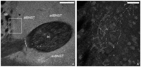
Figure 1. Confocal CB1 immunofluorescence in the anterior division of the rat BNST.
In A, intense CB1 labeling was in the anterodorsal (adBNST), anteroventral (avBNST) and anterolateral (alBNST) BNST around the anterior commissure (ac). In A′, an enlargement of the area framed in A shows CB1 immunoreactive varicose fibers in alBNST. Str: striatum. Scale bars: A: 200 µm; A′: 30 µm.

