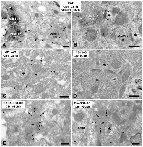
Figure 2. Immunolocalization of CB1 and vGluT1 in the rat anterolateral BNST (A, B).
Double labeling by combining a preembedding immunogold (CB1) and an immunoperoxidase (vGluT1) method for electron microscopy. vGluT1 immunoreactive presynaptic axonal terminals (vGluT1 + ter) making synapses with small dendrites (den) and dendritic spines (sp) localized CB1 immunoparticles at their perisynaptic and extrasynaptic membranes (white arrows). Observe in A, CB1 immunolabeling (black arrows) in a vGluT1 immunonegative synaptic bouton (vGluT1–ter); and in B, a vGluT1–synaptic terminal (vGluT1–ter) CB1 immunonegative. Small unmyelinated axons also showed CB1 immunoparticles (asterisks in A). The CB1 immunolabeled (arrows) synaptic terminals (ter) making synapses with small dendrites (den) and dendritic spines (sp) observed in wild-type mice (C) disappeared in CB1 −/− BNST tissue (D). CB1 immunolabeling (arrows) was detected in presynaptic boutons (ter) forming excitatory asymmetrical synapses with dendritic spines (sp) in the anterolateral BNST of GABA-CB1-KO mutant mice (E). Similarly, CB1 (arrows) was revealed in inhibitory-like axonal terminals (ter) making symmetrical synapses with small dendrites (den) in the anterolateral BNST of Glu-CB1-KO mutant mice (F). Scale bars: 0.4 µm.

