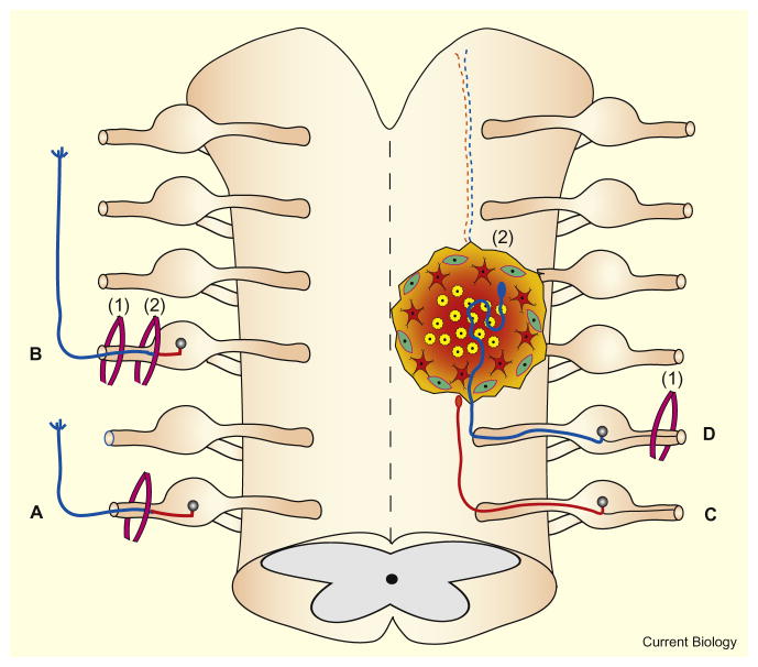
Figure 1. Conditioning lesions and regeneration in the CNS and PNS.
The left side of the figure depicts effects in the PNS and the right side depicts effects in the CNS. (A) After only a single PNS lesion, the rate of regeneration (blue) of severed axons is comparatively low. (B) After a conditioning lesion (1) in the PNS, the regeneration in the distal direction (blue) after a more centrally located test lesion (2) is significantly higher. (C) Regeneration in the CNS is minimal without conditioning as severed axons cannot cross scars. (D) A peripheral conditioning lesion (1) stimulates axonal regeneration within a lesion in the CNS (2).

