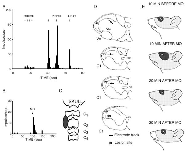
Fig 4.
Responses of a WDR neuron located in the C1 dorsal horn region. A shows a rate histogram depicting the neuron’s responses to 4 brushes, 2 noxious mechanical (pinch), and 1 noxious heat stimuli applied to its RF shown in E (top face figurine), and B illustrates its response to mustard oil (MO) injected into the deep cervical paraspinal tissues; the injection site shown in C was visualized by the extent of extravasated Evans blue dye. D indicates the reconstruction of the microelectrode tract (solid arrows) in Vc and C1 dorsal horn region; note the electrolytic lesion site (open arrow) in the bottom figurine. E indicates the neuron’s RF before and after MO was injected into the paraspinal tissues. Note the RF expansion for both tactile (solid area) and pinch (stippled region) components of the RF at 10 minutes after the MO injection and that the tactile RF had returned to its original size 20 minutes after the MO injection and that the pinch RF had returned to its original size 30 minutes after the MO injection.

