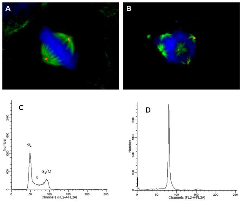
Figure 5.
Effects of 10i on mitotic spindles and cell cycle distribution. HeLa cells were treated with vehicle (A) or 0.50 µM 10i (B) for 18 h, fixed and cellular structures visualized by indirect immunofluorescence. Cellular microtubules were visualized with a β-tubulin antibody (green), centrosomes using a γ-tubulin antibody (red), and DNA using DAPI (blue). For the cell cycle distribution studies, MDA-MB-435 cells were treated for 18 h with vehicle (A) or 0.50 µM 10i then stained with Kirshan’s reagent and evaluation by flow cytometry.

