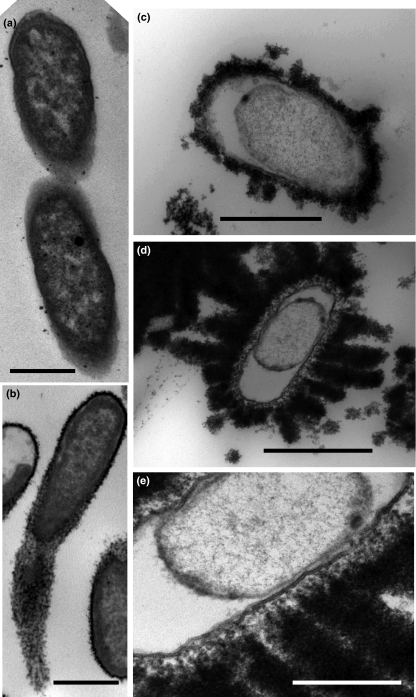
Figure 4.
Transmission electron microscopy observations of Klebsiella oxytoca BAS-10 (a) growing anaerobically in NaC medium; bar = 1 μm. (b) High electronic dense iron-binding capsular EPS of a cell growing anaerobically in FeC medium. Bar 1 μm was for (a) and (b) photos. (c) A short rod of Kl. oxytoca BAS-10 with detached inner membrane from wall and with very electron dense extrusion such as a bud of Fe(III)-EPS; bar = 0·5 μm. (d) A similar cell but with an overwhelmed production of Fe(III)-EPS like plumes; bar = 1 μm; (e) a detail of EPS emission in some spotted zones of envelope and formation of nano-vesicles out of outer membrane; bar = 0·5 μm.

