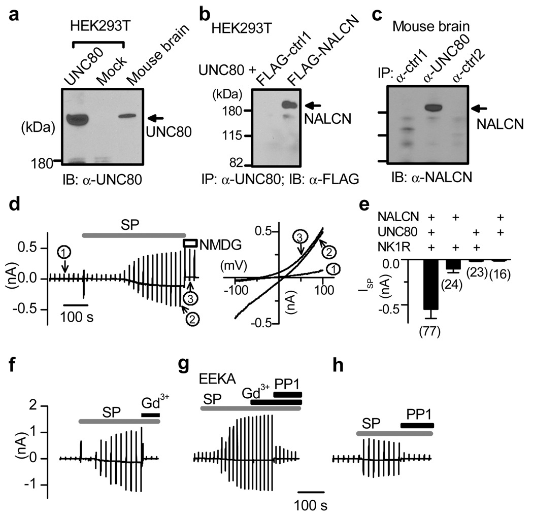
Figure 4. ISP reconstituted in HEK293T cells.
a, Immunoblot (IB) with lysates from transfected HEK293T cells and brain. b, c, Immunoprecipitation (IP) showing protein complex between mUNC80 and NALCN in HEK293T transfected as indicated (b) and in brain (c). Ctrl1 and ctrl2 were two unrelated proteins used as controls. Recordings in d–h were done using ramp protocols (Vh = −20 mV; −100 to +100 mV in 1 s, every 20 s). d, Recordings from a cell transfected with NK1R, mUNC80 and NALCN. Currents at three time points are expanded in the right panel (circled 1, before SP; 2, after SP; 3, after Na+ and K+ in the bath were replaced with NMDG). e, Summary of ISP sizes (at −100 mV) from cells transfected with combinations as indicated. f, g, Recordings showing that ISP was blocked by Gd3+ (f) but became resistant to Gd3+ when NALCN was replaced by the EEKA pore mutant (g). h, Inhibition of ISP by PP1 (20 µM). Error bars, mean ± s.e.m.

