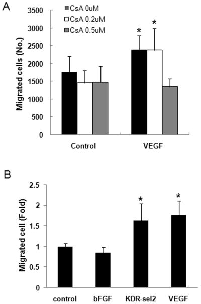
Fig. 2.
VEGF induced migration of valve endothelial cells in calcineurin- and KDR-dependent ways. A. Cell migration assay with HPVECs using Boyden chamber with polycarbonate membrane (8 μM pore size) was performed after VEGF were treated in a lower chamber and cell seeded onto upper chamber with or without CsA (0.2 or 0.5 μM). Migrated cells were counted after stained with hematoxylin and eosin (HE) and removed unmigrated cells. Significant differences from untreated control cells were noted as *p<0.01. B. Cells were treated with bFGF (10 ng/ml), KDR-selective variant VEGF (50 ng/ml), or VEGF (50 ng/ml) and migration assay using Boyden chamber was performed as described in A. Significant differences from untreated control cells were noted as *p<0.01. Data represent mean ± S.D. of a representative experiment (n=3), each performed in triplicate.

