
Figure 8.
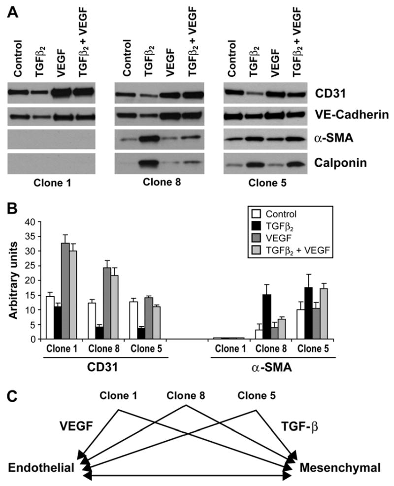
Endothelial/mesenchymal hierarchy in HPVEC clones. Clone 1 (left panel), Clone 8 (middle panel) and Clone 5 (right panel) were grown for 10 days in absence (control), 2ng/ml of TGFβ2 (TGFβ2), 10ng/ml VEGF (VEGF), or TGFβ2 and VEGF (TGFβ2+VEGF). Cell lysates were analyzed by western blot for the expression of CD31, VE-Cadherin, α-SMA and calponin. B. Quantitation of bands seen in A by densitometry. C. Schematic representation of hierarchy in plasticity of pulmonary valve progenitor cells.