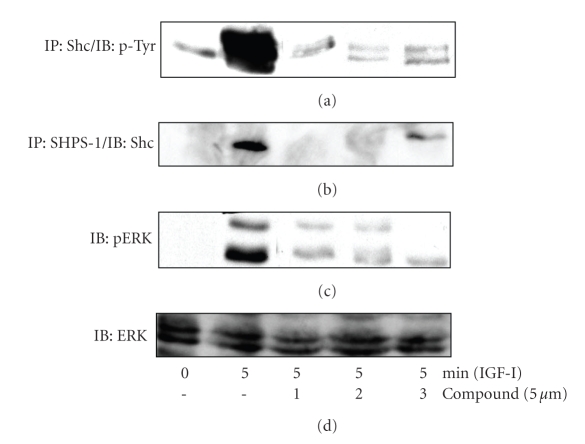
Figure 3.
Acceleration of IAP cleavage inhibits signaling in response to IGF-I. SMCs were grown to confluency in DMEM containing 25 mM glucose and then incubated overnight in HG-SFM. SMC were then incubated in HG-SFM, NG-SFM (5 mM) or HG-SFM containing the test compounds (μM) for 6 hours. Where indicated the cultures were treated with IGF-I (100 ng/mL) for 5 minutes prior to lysis. Cell lysates were then used for immunoprecipitation and proteins visualized by western immunoblotting. (a) Shc phosphorylation in response to IGF-I was determined by IP with an anti-Shc antibody and IB with an antiphosphotyrosine antibody (p-Tyr). (b) Shc association with SHPS-1 was determined following immunoprecipitation (IP) with an anti-SHPS-1 antibody and immunoblotting (IB) with an anti-Shc antibody. (c) MAPK activation was assessed by immunoblotting lysates directly with an antibody that specifically recognizes the phosphorylated forms of ERK1/2 (pERK). (d) To control for loading samples were immunoblotted with an anti-ERK antibody.

