Figure 2.
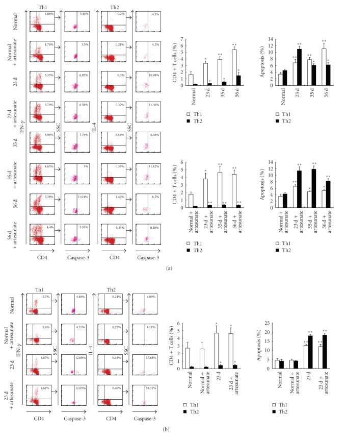
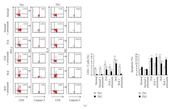
Kinetic analysis of Th1/Th2 cell polarization and apoptosis in mice infected with S. japonicum. In each experiment, twelve mice were percutaneously infected with S. japonicum and among these mice, six were orally administered artesunate dissolved in ddH2O as described in Materials and Methods. At 23, 35, and 56 days mice were sacrificed and single cell suspension of splenocytes were prepared. (a) CD4+ T cells were purified from splenocytes using MACS, surface stained with anti-CD4-FITC and then intracellularly stained with rabbit antimouse caspase-3 antibody or rabbit IgG isotype control antibody plus anti-IFN-γ-PE, anti-IL-4-PE, or isotype IgG control mAbs for FACS analysis. The splenocytes were restimulated in vitro with SWA (b) or SEA (c) for 36 hours, and then the CD4+ T cells were purified by MACS and stained with above antibodies prior to FACS analysis. The percentage of apoptotic cells in the FACS data was derived from the CD4+ and IFN-γ+, or CD4+ and IL- 4+ cells and gated on the caspase-3+ population. Data are expressed as the mean ± SD of 18 mice from three independent experiments. *P < .05; **P < .01. Left panels: One representative experiment of flow cytometric analysis with the average percentage of Th or apoptotic cells shown in the FACS data. Right panels: The statistical analysis of 18 mice from three independent experiments.


