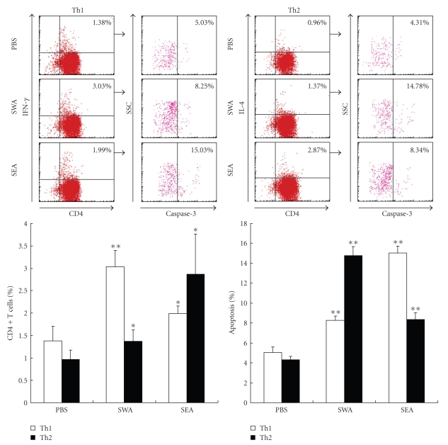
Figure 4.
In vitro induction of polarization and apoptosis in CD4+ T cells derived from normal mice with SWA and SEA. CD4+ T cells were purified from normal mice by MACS and preactivated overnight with anti-CD3 (2 μg/mL) and anti-CD28 (1 μg/mL) antibodies. Then the cells were stimulated with specific antigens of SEA, SWA or PBS alone for 36 hours at 37°C in 5% CO2, followed by staining with rabbit antimouse caspase-3 antibody or rabbit IgG isotype control antibody plus anti-CD4-FITC, anti-IFN-γ-PE anti-IL-4-PE mAbs, or isotype control antibodies prior to FACS analysis. The percentage of apoptotic cells in the FACS data was derived from the number of cells that were CD4+ and IFN-γ+ or CD4+ and IL-4+ and gated on the caspase-3+ population. Data are expressed as the mean ± SD of 18 mice from three independent experiments. *P < .05; **P < .01. Upper panels: One representative experiment of flow cytometric analysis with the average percentage of Th or apoptotic cells shown in the FACS data. Lower panels: The statistical analysis of 18 mice from three independent experiments.

