
Figure 4.
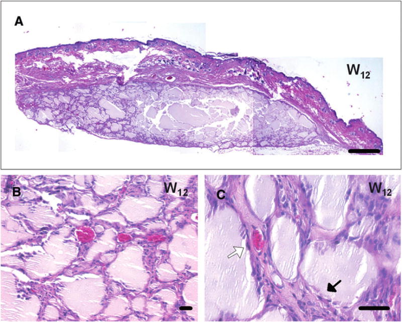
In vivo experiment: long-term histologic analyses. A through C) Histologic analyses with hematoxylin and eosin staining showed a complete disappearance of the inflammation process around silk gels at 12 weeks after grafting, with a partial fragmentation of the material. A) At this stage, a large piece of the silk material was detected. B and C) Fibroblasts and stromal tissues were found in the interstitial spaces between the silk-gel fragments (black arrow in C) associated with a complete vascularization (white arrow in C) of the area. Bar = 500 μm (A) and 50 μm (B and C).