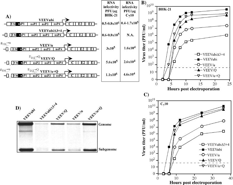
FIG. 4.
Replication of VEEVubiΔ3+4 variants, containing the mutations identified in the efficiently replicating VEEVrev1 pseudorevertant, in BHK-21 and C710 cells. (A) Schematic representation of VEEVubiΔ3+4 genomes with different mutations and infectivities of the in vitro-synthesized viral RNAs in the infectious center assay. Solid boxes indicate the nsP1 sequence containing clustered silent mutations. N.A. indicates “not applicable,” because the mutant was incapable of forming plaques in C710 cells. (B and C) Single-step virus growth curves after electroporation of 3 μg of in vitro- synthesized RNAs into BHK-21 or C710 cells. At the indicated times, the medium was replaced and virus titers were determined in BHK-21 cells, as described in Materials and Methods. The dashed lines indicate the limit of detection. (D) Synthesis of virus-specific RNAs in the transfected BHK-21 cells. At 2 h post transfection, viral RNAs were metabolically labeled with [3H]uridine for 6h, as described in Materials and Methods, and analyzed by agarose gel electrophoresis. Positions of the genomic and subgenomic RNAs are indicated.

