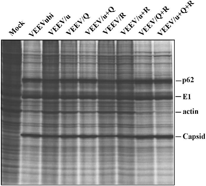
FIG. 7.
Protein synthesis in BHK-21 cells infected with different variants of VEEVubiΔ3+4. Cells were infected with the viruses indicated at an MOI of 10 PFU/cell. At 20 h post infection, they were labeled with [35S]methionine and analyzed in a sodium dodecyl sulfate-10% polyacrylamide gel as described in Materials and Methods. Positions of VEEV-specific structural proteins and actin are indicated.

