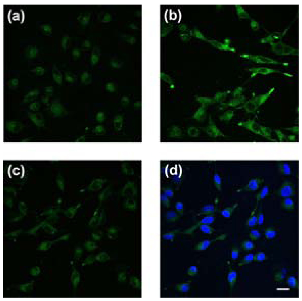
Figure 2.
Live-cell imaging of intracellular Ni2+ levels by confocal microscopy. (a) Control A549 cells incubated with a 1:1 mixture of 10 µM NS1-AM and F-127 Pluronic acid for 35 min at 37 °C. (b) Cells supplemented with 1 mM NiCl2 in the growth medium for 18 h at 37 °C and stained with 10 µM NS1-AM and F-127 Pluronic acid for 35 min at 37 °C. (c) NS1-loaded, 1 mM Ni2+-supplemented cells treated with 1 mM of the divalent metal chelator TPEN for 1 min at 25 °C. (D) NS1-loaded, 1 mM Ni2+-supplemented cells treated with 1 mM TPEN, stained with 5 µM Hoescht-3342 to show cell viability. Scale bar = 20 µm.

