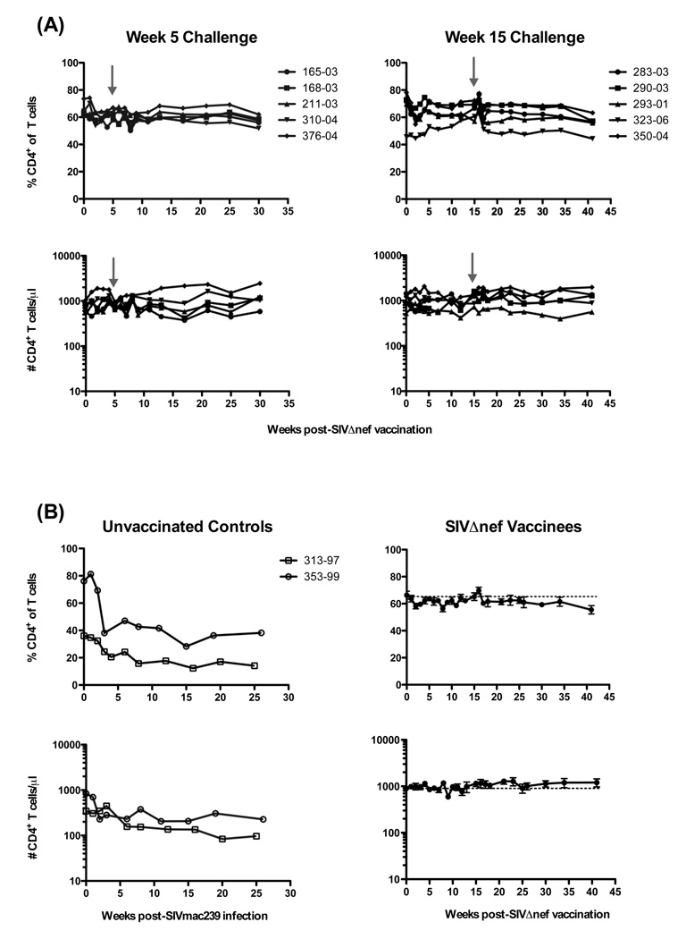
Figure 3.
Longitudinal analysis of circulating CD4+ T cells in SIVΔnef-vaccinated and SIVmac239-infected rhesus macaques. (A) Frequencies (upper panels) and absolute numbers (lower panels) of circulating CD4+CD3+ lymphocytes are shown for SIVΔnef-vaccinated macaques challenged intravenously with SIVmac239 at either 5 or 15 weeks post-vaccination; grey arrows indicate 5 and 15 week challenges, respectively. (B) Frequencies and absolute numbers of CD4+CD3+ lymphocytes in two unvaccinated controls challenged with SIVmac239 (left panels) and means ± SEM of SIVΔnef-vaccinated macaques (right panels) combined from (A). Dashed horizontal lines indicate means at day of vaccination (day 0).

