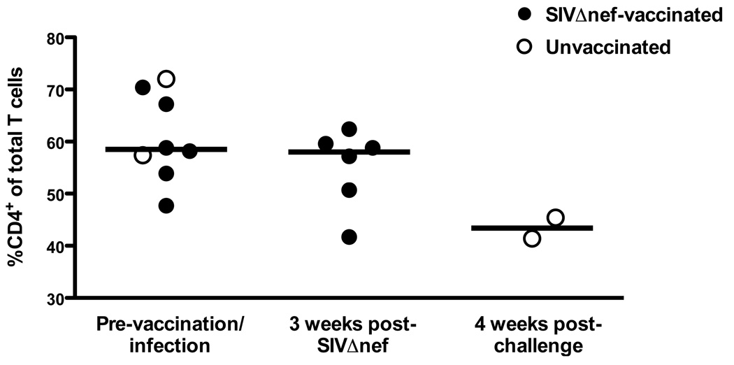
Figure 5.
CD4+ T cell frequencies in the rectal mucosa of SIV-naïve, SIVΔnef-vaccinated, and SIVmac239-infected rhesus macaques. Percentages of CD4+ T cells are shown as frequencies among total T cells in lymphocytes isolated from rectal biopsies performed at one week prior to and three weeks after SIVΔnef vaccination and four weeks after SIVmac239 challenge (weeks 5 and 15 challenge groups are combined). Bars indicate medians of two to eight animals.

