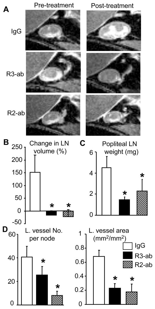
Figure 3.
VEGFR-2 and VEGFR-3 neutralizing antibodies prevent TNF-induced PLN enlargement and lymphatic vessel formation. TNF-Tg mice (2.5-month-old, N=5/group) received CE-MRI of ankle and knee joints to obtain baseline value of synovial and PLN volume. Animals were then treated with VEGFR-2 or VEGFR-3 antibodies, or IgG (0.8 mg/mouse, i.p. 2/week) for 8 weeks. The post-treatment CE-MRI were performed one day before sacrifice. (A) Representative 2D CE-MRI images show a marked increase in IgG treated PLN volume over 8 weeks, compared to the lack of changes in PLN from antibody treated animals. (B) The % change in PLN volume from baseline is presented as mean ± SD for each treatment. (C) The PLN weights after 8-weeks are presented as mean ± SD for each treatment. (D) Histomorphometry of lymphatic vessel number and area in anti-LYVE-1 antibody-stained sections. The data are mean ± SD for each treatment.*p<0.05 vs. IgG.

