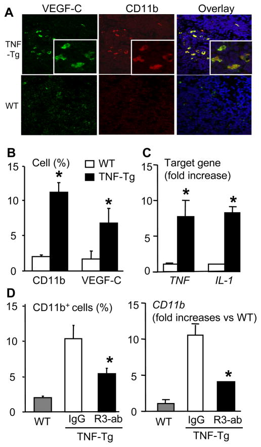
Figure 6.
VEGFR-3 blockade decreases VEGF-C-expressing CD11b+ cells in lymph nodes of TNF-Tg mice. (A) PLNs from TNF-Tg mice (4–6-month-old) and WT littermates were double immuno-stained with PE-anti-CD11b and FITC-anti-VEGF-C antibodies. Representative pictures taken at 40x indicate the co-localization of CD11b and VEGF-C proteins. (B) Histomorphometry was performed to determine the percentage of CD11b+ or VEGF-C+ cells in the PLNs. The values are the mean ± SD of 4 fields from 4 individual mice per genotype (*p<0.05 vs. WT). (C) Total RNA was extracted from pooled (n= 3–4) PLNs and the expression levels of TNF and IL-1 were determined by real-time RT-PCR standardized to a β-actin control. The data are presented as the mean fold-increase ± SD vs. the mean WT expression (*p<0.05 vs. WT). (D) PLNs from IgG- and VEGFR-3 neutralizing antibody-treated TNF-Tg mice were stained with anti-CD11b antibody, and the percentage of CD11b+ cells was calculated by dividing the number of CD11b+ cells by the total number of cells (left panel). The expression levels of CD11b mRNA were determined by real time RT-PCR (right panel). The data are presented as the mean fold-increase ± SD vs. the mean IgG expression. *p<0.05 vs. IgG treated mice.

