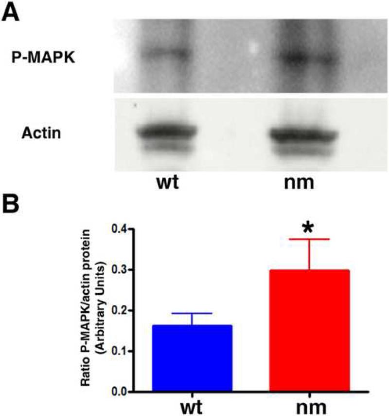
Figure 10. Phosphorylated MAPK is increased in E6 nm cartilage.
A. Immunobloting analysis showing levels of phosphorylated MAPK (Erk), and β-actin as a loading control, in E6 wt and nm cartilage lysates. B. Relative levels of phosphorylated MAPK to β-actin were quantified in three independent experiments. Data was evaluated for statistical significance using the Student's t-test. *< 0.04.

