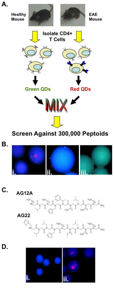
Figure 1. Identification of putative autoreactive T cell binding peptoids using a bicolor on-bead screening protocol.

(A) Schematic representation of the peptoid screening protocol. CD4+ T cells isolated from EAE mice have an increased frequency of Vα2.3/Vβ8.2 MBP Ac1-11 specific T cell receptors compared to wild type healthy control littermates. Following isolation, T cells were differentially labeled with green and red quantum dots and screened against a bead-displayed peptoid library. Peptoid beads binding only cells from EAE mice were selected and sequenced. (B) Fluorescent microscopic images of peptoid beads after screening and washing (100X magnification; DAPI filter). i.and ii. Photographs depicting the 2 putative hit peptoid beads bound to CD4+ T cells from EAE mice (red stained cells). iii. Photograph depicting peptoid beads binding to CD4+ T cells from healthy mice and EAE mice. (C) Chemical structures of the two hits identified in the screen. (D) Fluorescent microscopic images of tentagel beads displaying AG12A bound to autoreactive T cells. i. CD4+ T cells from B10.PL wildtype control mice do not bind AG12A peptoid beads. ii. CD4+ T cells from Vα2.3/Vβ8.2 MBP Ac1-11 TCR transgenic mice bind to AG12A peptoid beads.
