
Figure 2.
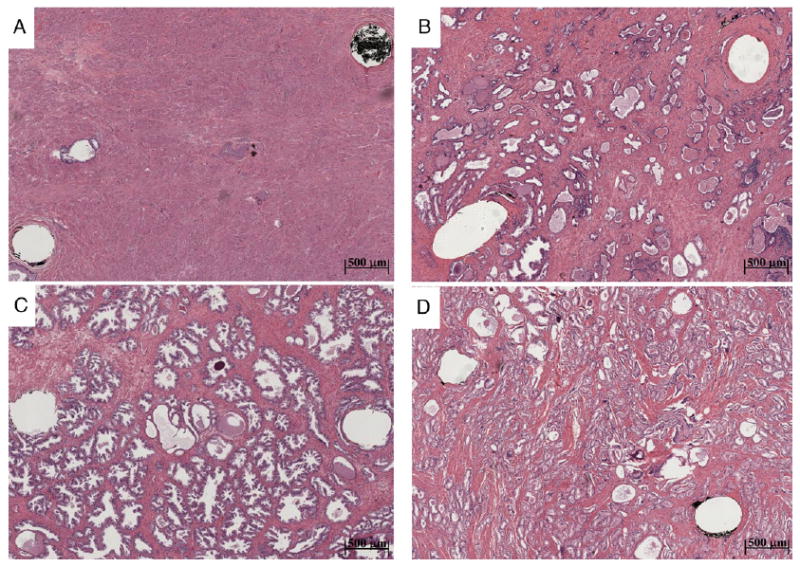
Photomicrographs show 100% Str (A), Gl (B), BPH (C) and ACa (D) tissue types. Probed region corresponds to area between 2 visible pinholes remaining after tissue fixation and slide preparation. Note decreased Str content in more glandular tissue types, which we hypothesize largely influences tissue conductivity. Variability in gland size and configuration likely influences permittivity associated with different tissue types.