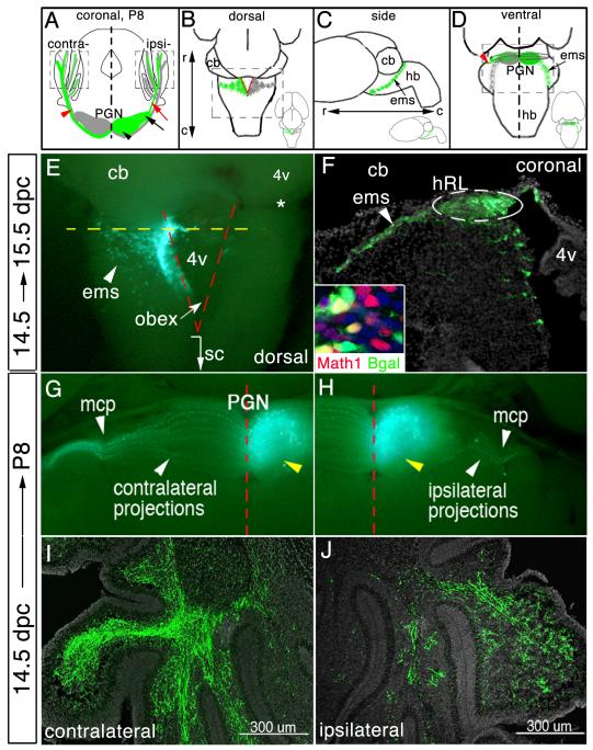
Fig.1.
Visualizing development of a single lobe of the bilateral PGN. (A) Schematic of oblique coronal brain section. The PGN is comprised of rostromedial (black arrowhead) and caudolateral (black arrow) neuronal subpopulations on each side of the brain stem. Green marking represents eGFP/nßgal-transfected neurons and their axons. Most PGN neurons extend axons across brainstem midline toward the contralateral cerebellum (red arrowhead), whereas few project away from the midline toward the ipsilateral cerebellum (red arrow). Dashed boxes, “contra” and “ipsi”, identify area of cerebellar images in (I) and (J), respectively. (B-D) Whole brain schematics. Insets, low-power images of brains. PGN precursor cells emerge from the hRL (red lines) (B) and travel ventrally forming the extramural migratory stream (ems) (C). Postmitotic hRL precursor cells aggregate adjacent to the ventral brainstem midline to form the PGN, extending axons across or away from the midline (D). Dashed boxes in (B) and (D) identify area of images in (E), and (G,H), respectively. (E) Whole brain at 15.5 dpc that was electroporated at 14.5 dpc. eGFP-transfected mitotic cells within the hRL and postmitotic cells within the ems. Dashed yellow line in (E) identifies the ideal axial level of the coronal section in (F). Dashed red lines in (E) demarcate location of the left and right side of the hRL. eGFP+ cells form the ems (arrowhead) (E and F). (F) Immunodetection of eGFP on a high magnification coronal section shows transfected progenitor cells within one side of the hRL and their postmitotic progeny cells within the ems. Inset in (F), co-immunodetection for nßgal and Math1. (G,H) Ventral view of P8 brains that were electroporated at 14.5 dpc. eGFP fluorescence shows labeled cells in the ipsilateral lobe of the PGN (yellow arrowhead) and their contralateral (G) and ipsilateral (H) MF projections. Immunodetection of eGFP shows eGFP+ MF axons throughout the granule cell layer of the contralateral (I) and ipsilateral (J) cerebellum. DAPI staining to highlight cell bodies is shown in gray. r, rostral; c, caudal; cb, cerebellum; sc, spinal cord; 4v, fourth ventricle; mcp, medial cerebellar peduncle.

