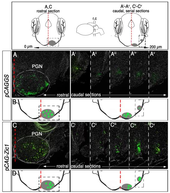
Fig. 4.
Zic1 overexpression in cells of the precerebellar MF lineage leads to a redistribution of neurons to caudolateral territories. Above, schematics of coronal sections along the rostrocaudal axis of the PGN at P8. Inset (center) depicts side view of the brain, and black dashed lines indicate idealized axial levels of coronal sections to the left and right. Distance between rostral and caudal sections is ~200 μm; PGN is in gray. Red dashed line demarcates brainstem midline. Boxed area in the rostral schematic identifies neurons in rostromedial PGN in (A) and (C). Boxed area in the caudal schematic identifies region in caudolateral PGN (Ai-Av) and (Ci-Cv). (A and Ai-v) Immunodetection for nßgal highlights cells co-transfected with control vector (pCAGGS) and reporter vectors. Coronal section of the rostral PGN of a P8 animal that was electroporated at 14.5 dpc (B). Five serial sections of the caudal PGN (A-Av). (B) Summary schematic of labeled cells within the PGN of control animals. (C and Ci-v) Immunodetection for nßgal shows cells co-transfected with pCAG-Zic1 and reporter vectors. Coronal section of the rostral PGN (C). Five serial sections of the caudal PGN (C-Cv). A significantly greater proportion of nßgal+ cells (transfected with pCAG-Zic1) reside in caudolateral PGN as compared to control animals in (Ai-Av) (student’s t-test, p<0.0001). (D) Summary schematic of labeled cells that have shifted to caudolateral aspects of the PGN in Zic1 overexpression animals.

