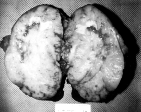
Fig. 2.
Gross examination of the resected spleen of Case 1. The exposed cut surface shows a bulging, well-demarcated grayish-yellow nodular solid mass, measuring 12 cm in the largest dimension. Multifocal grayish-yellow necrotic foci are present. The splenic parenchyma is nearly replaced by this lesion.

