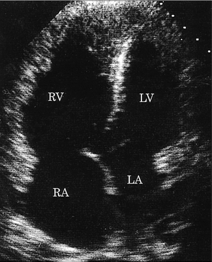
Fig. 2.
Apical four-chamber view of the echocardiogram demonstrated marked dilatation of the right ventricle and right atrium. The interventricular septum and the atrial septum were deviated towards the left ventricle and left atrium respectively. RV, right ventricle; RA, right atrium; LV, left ventricle; LA, left atrium.

