
Figure 1.
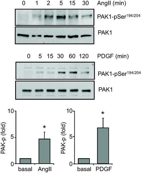
Phosphorylation of PAK1 by AngII and PDGF-BB. VSMCs were stimulated with 100 nmol/L AngII or 100 ng/mL PDGF-BB for the indicated time periods. The cell lysates were immunoblotted with a phospho-selective antibody, which detects PAK1-pSer194/204 phosphorylation, and with anti-PAK1 antibody. The bar graphs show quantification of the PAK1 phosphorylation by densitometry at 5 min and 30 min induced by AngII and PDGF-BB, respectively. Data are mean±SEM of 3 experiments. *p<0.05 compared to the basal control.