
Figure 4.
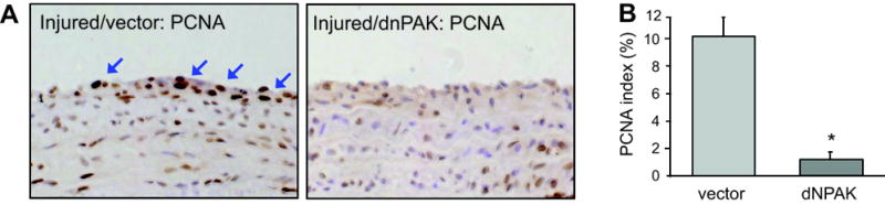
Histological analysis of cell proliferation in arterial cross-sections obtained after balloon injury. Arterial sections obtained on day 14 after injury with infection of adenovirus encoding GFP or dnPAK1 were stained with PCNA antibody (x400 magnification). Representative sections (each from 4 sections, x400 magnification) are shown. A, Cells were considered positive for PCNA expression only in the presence of an intense brown staining of the nucleus. The arrows indicate representative PCNA positive cells in the neointima lesion. B, The number of PCNA-positive cells was expressed as a percentage of the total cell number in the neointima (PCNA index). Data are mean±SEM of 4 sections. *p<0.05 compared to the GFP adenovirus-infected control.