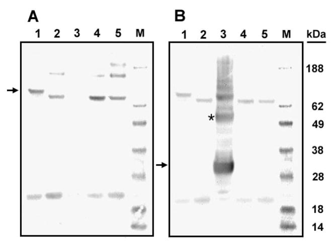
Figure 3.
Western blot analyses of one-chain carboxylase and one- and two-chain L368/372P carboxylase and PNGase F-treated forms of these carboxylases. Microsomes from cells expressing either wild-type carboxylase or the carboxylase L368/372P were analyzed by SDS–PAGE under nonreduced conditions and transferred to a polyvinylidene difluoride membrane. Protein bands were visualized using the antibody to the C-terminal carboxylase peptide (A) or the antibody to the N-terminal peptide (B) employing ECL reagents. One-chain wild-type carboxylase (lane 1 in panels A and B), one-chain L368/372P (lane 2 in both panels), two-chain L368/372P (lane 3 in both panels), PNGase F-treated wild-type carboxylase (lane 4 in both panels), and PNGase F-treated one-chain L368/372P (lane 5 in both panels). One-chain carboxylase (A) and the N-terminal peptide (B) are indicated by arrows. The dimer of the N-terminal peptide (B) is indicated by an asterisk.

