
Fig. 1.
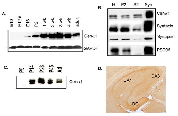
Developmental expression and localization of endogenous centaurin α1 in the rodent brain. (A) Immunoblot analysis of centaurin α1 protein from lysates (25 μg total lysate per lane) from whole brain at different developmental stages from E10 to P42 (=adult). (B) Immunoblot analysis of centaurin α1 and synaptic markers in fractionated P23 whole rat brain. H, whole brain homogenate; P2, crude microsomes and synaptosomes; S2, supernatant; Syn, purified synaptosomes; 20 μg of each fraction was loaded per lane. (C) Immunoblot analysis of centaurin α1 in the synaptosome fraction prepared from the developing brain. 10 μg of each fraction was loaded per lane. (D) Immunohistochemistry with anti-centaurin α1 antibody in hippocampus from P28 rat. DG, dentate gyrus.