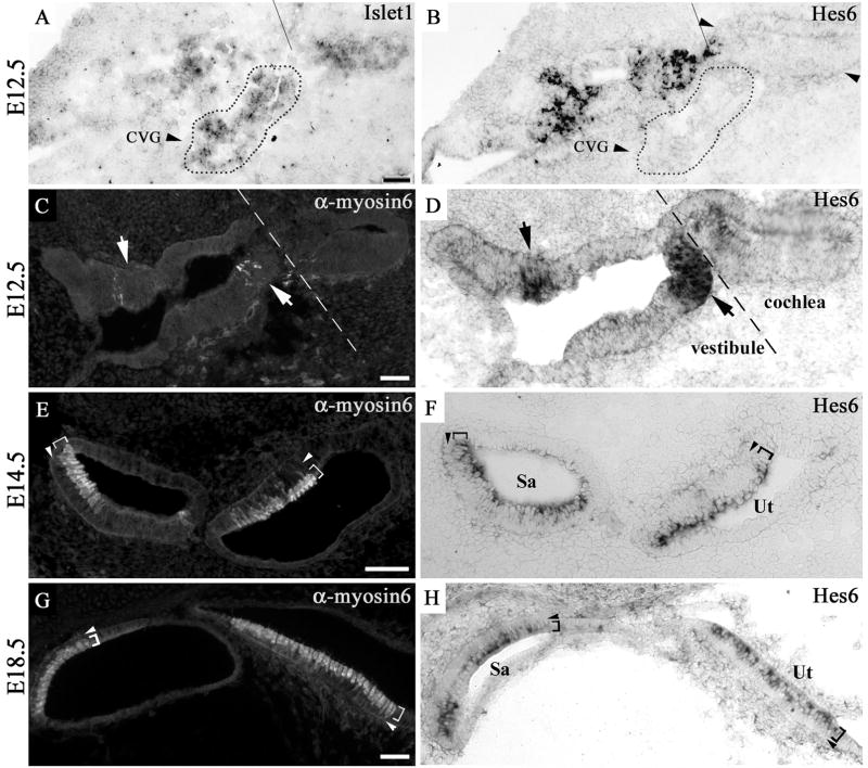
Fig. 1.
Hes6 expression in the vestibular sensory organs. The adjacent inner ear sections from an E12.5 embryo (A–B and C–D), and the adjacent vestibular sections from E14.5 (E–F) and E18.5 (G–H) embryos were probed for Islet1 message (A) or Hes6 message (B, D, F, H) or stained for myosin6 (C, E, G). Islet1 is expressed in the floor of the cochlear duct and in the cochleovestibular ganglion (A, marked by dotted outline), while Hes6 message is not detected in the corresponding regions (B). The sensory regions of the vestibule at E12.5 were recognized by the expression of the hair cell-specific marker myosin6 (C, indicated by arrows). The expression of Hes6 is detectable in the otocyst by E12.5 in the same region in an adjacent section (D, indicated by arrows). The vestibular and cochlear regions of the inner ear at E12.5 are indicated, and the dashed line approximates where the two regions meet (A–D). By E14.5, the expression of myosin6 (E) and Hes6 (F) is limited to the luminal hair cell layer (E–F, indicated by brackets) of the vestibular sensory organs. The expression of Hes6 in the hair cells in the vestibule persists at E18.5 (G–H). The arrowheads (E–H) mark the supporting cell layer underneath the luminal hair cell layer. CVG: Cochleovestibular ganglion. Sa: Saccule. Ut: Utricle. Scale bars: 50 μm.

