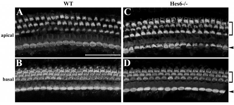
Fig. 6.
Inner ear sensory organs are normal in Hes6 null animals. The cochleae from wild type control (A–B) and Hes6 null (C–D) littermates at 6 month old were stained for myosin6. Both the apical (A, C) and the basal (B, D) regions are shown. Arrows and brackets indicate the inner and outer hair cells, respectively. The organ of Corti from the Hes6 null animals consists of the normal three rows of outer and one row of inner hair cells. No abnormality was observed in Hes6 null animals. Scale: 50 μm.

