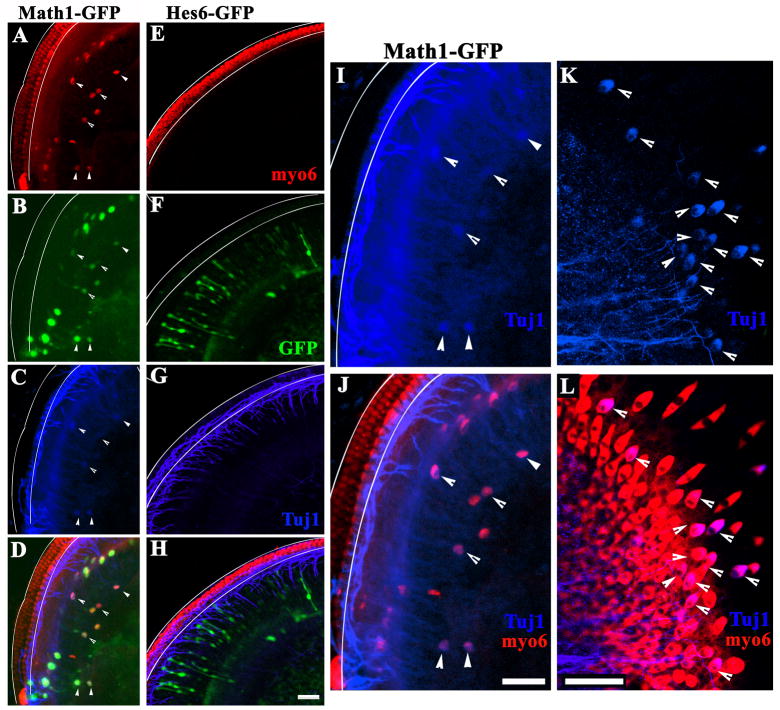
Fig. 7.
Hes6 is not sufficient to induce sensori- or neuronal marker expression in the cochlear epithelium in vitro. Neonatal rat cochlear epithelia were electroporated with Math1-IRESGFP (A–D) or Hes6-IRESGFP (E–H) expression vectors, and cultured for 6 days in vitro. The cultures were stained for both myosin6 (A, E, myo6), and tubulin β–III (C, G, blue). Transfected cells express GFP (B, F, green) in Math1 (B) or Hes6 (F) transfected cells. The overlays of the myosin6 (red), GFP (green), and tubulin β-III (blue) for Math1-GFP and Hes6-GFP transfected cultures were shown in (D) and (H), respectively. The pair of solid lines (A–J) outline the organ of Corti where endogenous hair cells are stereotypically arrayed. Arrows (A–D) indicate Math1 transfected cells positive for both myosin6 and tubulin β-III. The images in C and D at a higher resolution are shown in I and J, respectively, to better visualize the cells that were positive for both myosin6 (J, red) and tubulin β-III (I–J, blue). Hair cells positive for tubulin β-III were also seen in utricle cultures (K–L, blue). Scale bars (A–H, I–J, K–L): 50 μm.

