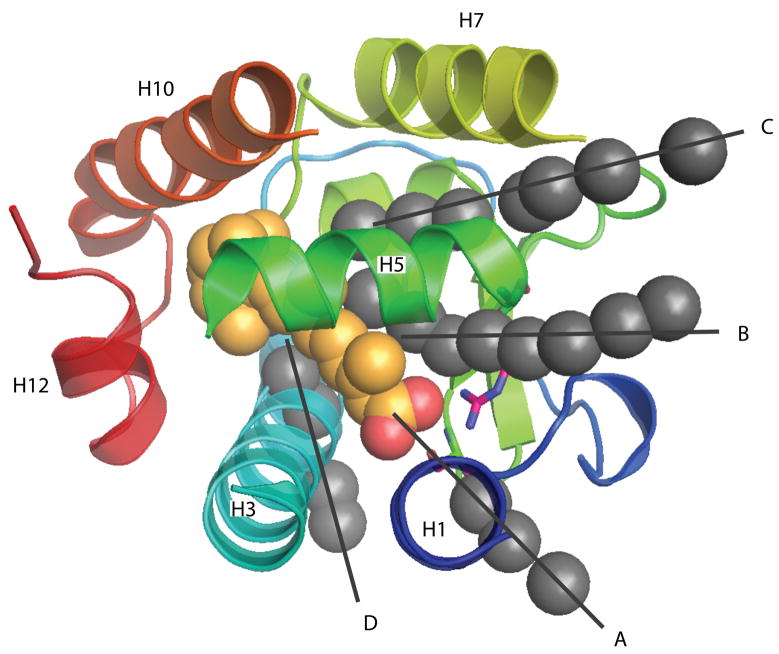
Figure 4.
Four different (A-D) ligand escape pathways (shown as grey spheres along black guiding lines) identified using Random Acceleration Molecular Dynamics35 in the ligand binding domain of the retinoic acid receptor. Helices are shown as ribbons, and the retinoic acid ligand in the bound initial state is shown as red and gold spheres. (From Carlsson et al.35).

