
Figure 8.


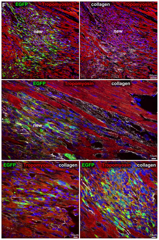
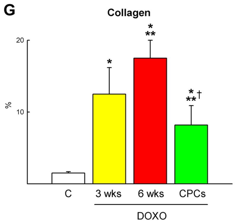
Myocardial regeneration and DOXO-induced cardiomyopathy. A, Low-power view of transverse sections of the LV wall of DOXO-treated rats 3 weeks after CPC injection. Arrows define areas of myocardial regeneration (green). Newly formed myocytes express EGFP (green) and α-SA (red). B, The area of myocardial regeneration included in the rectangle is shown at higher magnification in the adjacent panel. Small myocytes in the cluster express EGFP (green) and α-SA (red). C, Newly formed EGFP-positive (green) myocytes are labeled by BrdU (white) and express α-SA (red). D, Number of newly formed capillaries and arterioles. E, Foci of collagen (yellow) accumulation at times surrounding small vessels. F, Regenerated myocytes (new) are positive for EGFP (green) and tropomyosin (red) and replace areas of fibrosis (collagen, white). G, Fraction of collagen within the myocardium. *,**,† P <0.05 vs. c, 3 wks and 6 wks, respectively.