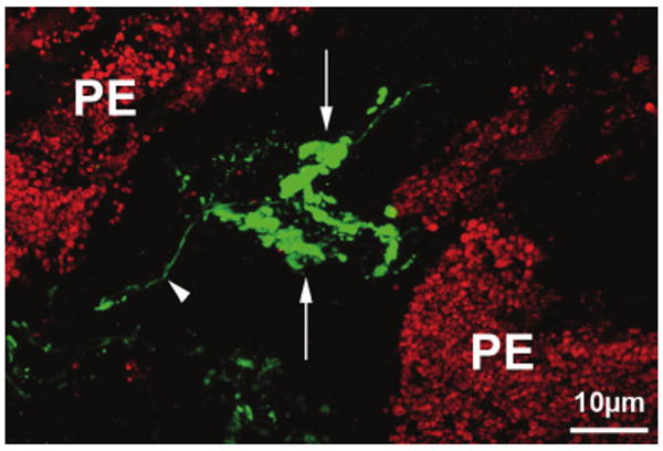
Figure 4.

Confocal all-in-focus image of a frozen tangential section through the valley of a ciliary process within the anterior pars plicata, immuno-labeled for calretinin (green). Complex Ruffini ending-like arborizations (arrows) of a calretinin-IR fiber (arrowhead) can be seen next to cells of the pigmented epithelium (PE).
