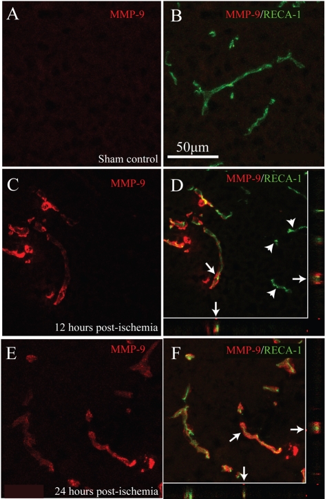
Figure 4. Microvascular MMP-9 expression is induced by ischaemic SCI.
In control ventral grey matter, no detectable MMP-9 immunoreactivity is present in smvECs (A and B). By 12 h post-ischaemia, significant MMP-9-immunoreactvity is observed in affected spinal microvessels (C and D), although a subset of microvessels does not express MMP-9 (D; arrowheads). This increase in MMP-9 immunoreactivity is maintained 24 h post-ischaemia (E and F), with all microvessels in affected tissue expressing detectable levels of MMP-9. Confocal analysis confirms co-localization of RECA-1 and MMP-9 in the xz and yz planes (D and F; arrows). Scale bar = 50 μm (A–F).

