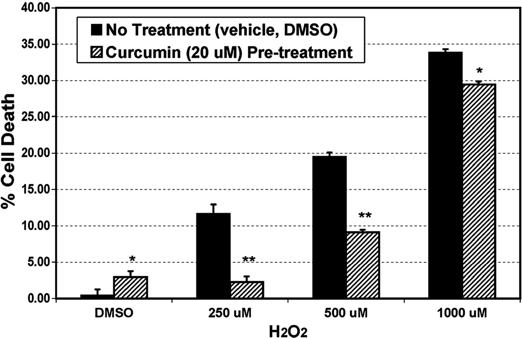
FIGURE 5.
Cell viability assay. Freshly grown 661W cells in 10 cm plate were pre-treated in situ with 20 µM curcumin or with the vehicle (DMSO) for 3h. After thorough washing the cells were exposed to different doses of H2O2 for 3h. Cell death was indirectly measured by measuring the release of lactate dehydrogenase (LDH) in the media in which a plate containing 2% Triton served as 100% cell death (n = 4 plate × 4 replication assay). *P < 0.01, and **P < 0.001, by Student’s t-test.

