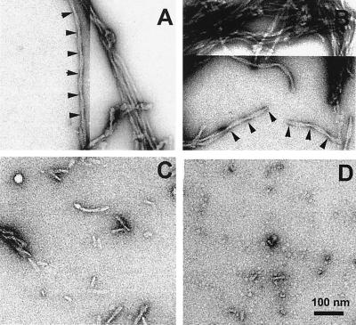
Figure 2.
Electron micrographs of PHFs formed by spontaneous or seeded assembly. (A) PHFs formed by spontaneous assembly of tau construct K19 (20 μM) after dimerization in the presence of poly-Glu. (B) PHFs formed by seeded assembly of K19 dimers (with poly-Glu). The concentration of seeds corresponds to 1 μM K19 monomers. The estimated seed number concentration is ≈2–20 nM (based on apparent PHF fragment lengths of 10–80 nm and a mass density of ≈60–70 kDa/nm, data not shown). (C and D) Seeds made by sonication for 2 or 5 min of filaments shown in A. Arrowheads indicate crossover points of the two strands of a PHF, spaced about 80 nm. The variations in stain accumulation were compensated by adjusting the contrast by image processing to make the filament structure more visible. (Bar = 100 nm.)

