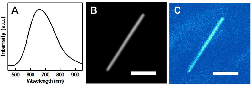
Figure 4.
(A) Photoluminescence spectrum of porous silicon nanowires obtained with 60 min etching in a solution with 0.30 M H2O2; (B) Optical micrograph of a single porous silicon nanowires; and (C) Confocal photoluminescence image of the same single porous silicon nanowire. The scale bar in (B) and (C) is 3 µm.

