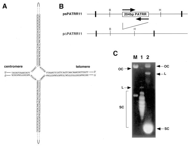
Fig. 1. Agarose gel electrophoresis of the PATRR plasmid.
A, putative cruciform structure of the PATRR of chromosome 11. B, schematic representation of psPATRR11 and pΔPATRR11. The box indicates the inverted repeat region, whereas each arrow indicates the repeat unit. pΔPATRR11 deletes almost the entire 204-bp PATRR region. Bold vertical lines indicate the cloning sites of the plasmids. The thin vertical lines indicate restriction sites for the following enzymes: BamHI (B) and HincII (H). C, agarose gel electrophoresis of psPATRR11 and pΔPATRR11. Lane M, molecular size marker; lane 1, psPATRR11; lane 2, pΔPATRR11. OC, open circle; L, linear; SC, supercoiled circle.

