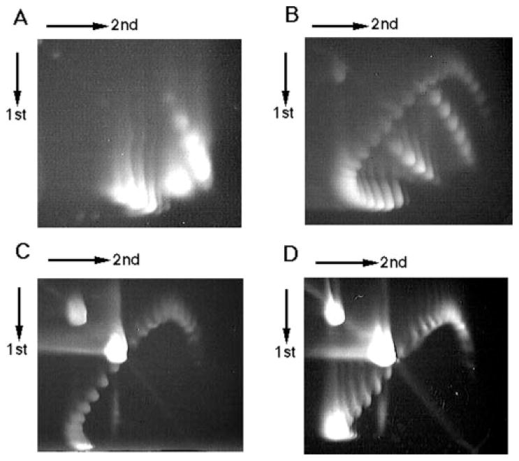
FIG. 3. Two-dimensional gel electrophoresis of the PATRR plasmid.
A, psPATRR11 prepared by the alkaline lysis method. B, psPATRR11 treated with topoisomerase I in the presence of various amounts of ethidium bromide. C, topoisomerase I-treated psPATRR11 digested with T7 endonuclease. D, topoisomerase I-treated psPATRR11 digested with S1 nuclease. Two downward-sloping curves originating from cruciform extrusion are observed at the lower right side on both the A and the B gels, but neither are observed in the C nor in the D gel. Spots at the upper left side on the gels originate from open circular nicked plasmids, whereas spots near the center of the gels are from nuclease-cleaved linear plasmids.

