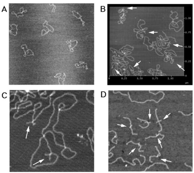
FIG. 7. AFM image of the PATRR plasmid.
A, an AFM image of psPATRR11 prepared by the Triton lysis method. B, an AFM image of the psPATRR11 after incubation at room temperature. C, another image of psPATRR11 at a higher magnification. D, an AFM image of psPATRR11 after DNA cross-linking followed by restriction digestion. The arrows indicate the cruciform extrusion. Longer fragments without cruciform structure may originate from the cloning vector. The scale bar is indicated in μm.

