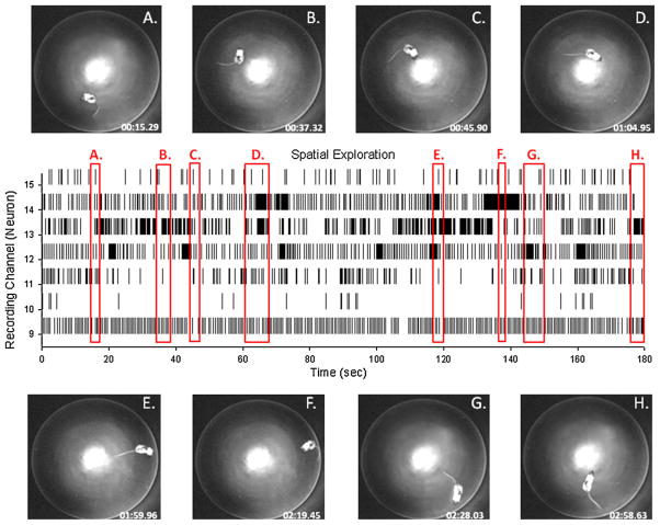
Fig. 7.
Implementation of UBAT in open field test for spatial firing correlates of hippocampal neurons. (A–H) Overhead photos of rat in different spatial locations in circular open field recording chamber. Center display shows firing during spatial exploration via continuous stripchart records from 7 different neurons (channels 9–15) with red rectangles indicating entry and exits from locations labeled in overhead photos. UBAT transmission and receipt of signal utilized the same monitoring station as the DNMS task in Fig. 6; however, the rat and apparatus were located in a different room in the laboratory, indicating lack of necessity of line of sight orientation between the UBAT and monitoring station.

