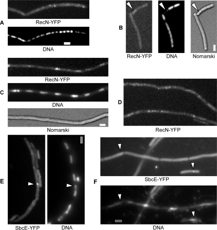
Figure 6.
Localization of RecN-YFP or SbcE-YFP in dnaBts cells. (A–B) RecN-YFP foci in cells shifted to 42°C (non-permissive temperature) for 2 h, white arrowheads in (B) indicate cell with obvious segregation defect lacking any RecN-YFP foci. (C–D) RecN-YFP foci in cells shifted to 42°C for 60 min, after addition of MMC for further 60 min. (E) SbcE-YFP foci (indicated by white arrowheads) in cells shifted to 42°C for 2 h. (F) SbcE-YFP foci (indicated by white arrowheads) in cells shifted to 42°C for 60 min, after addition of MMC for further 60 min. Grey bars 2 µm.

