Figure 6.
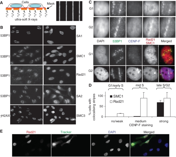
Recruitment of cohesin to ionizing radiation-induced DNA double strand breaks. (A) Schematic representation of micro-irradiation setup (left) and bright field image of the grid with 1 μm wide gaps/9 μm wide shielding used for generating defined patterns of DNA damage within the nucleus (right). (B) HeLa cells were grown on Mylar foil and irradiated with soft X-rays. After 1 h incubation, cells were pre-extracted with 0.1% Triton X-100, fixed and stained with the indicated antibodies. Each image is 125 μm wide. (C) As in (B) but cells were furthermore stained with CENP-F to determine the cell cycle phase. Each image is 15 μm wide. (D) Quantitative analysis of Rad21 and SMC1 stripe formation. Cells were divided into three groups of no or weak, medium and strong CENP-F staining. Microbeam tracks were predicted using the Rad21 or SMC1 signal, respectively, and then tested for co-localization with the 53BP1 signal. (E) Immunofluorescence microscopic analysis of Rad21 depletion. Control siRNA-transfected cells labelled with CellTrackerTM Green CMFDA (Invitrogen) were mixed with Rad21 siRNA-treated cells and stained for Rad21.

