
FIGURE 4.
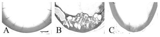
Vacuole formation in the TGF-β1/Smad3 lenses. The posterior lens cortex of wild-type (A), TGF-β1/Smad3+/+ (B), and TGF-β1/Smad3-/- (C) lenses are shown. The expression of TGF-β1 induced nucleation and vacuole formation in the TGF-β1/Smad3+/+ lenses. Both the wild-type and TGF-β/Smad3-/- lenses showed normal morphology of the posterior lens cortex. Scale bar, 200 μm.