
FIGURE 6.
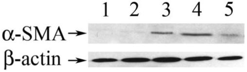
α-Smooth muscle actin protein expression. Western blot analysis using an anti-α-SMA antibody for the detection of α-SMA (42 kDa) protein expression in lens extracts. The membrane was stripped and reprobed for β-actin, which served as a loading control. Lanes 1 to 5: α-SMA and β-actin protein expression in wild-type, Smad3-/-, TGF-β1/Smad3+/-, TGF-β1/Smad3+/+, and TGFβ1/Smad3-/- lenses, respectively. The β-actin signal shows that equal amounts of protein were loaded in all lanes. TGF-β1/Smad3+/+, TGF-β1/Smad3+/-, and TGFβ1/Smad3-/- lenses showed expression of α-SMA, whereas wild-type and Smad3-/- lenses did not. The TGFβ1/Smad3-/- lenses showed reduced levels of α-SMA compared with both the TGF-β1/Smad3+/+ and TGF-β1/Smad3+/- lenses.