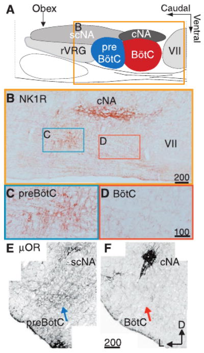
Fig. 2.

NK1R- and μOR-expressing neurons are limited to and define the preBötC. (A) Cartoon indicating approximate location and boundaries of ventral respiratory column regions and adjacent ambiguual motoneuron pools in sagittal section. (B) NK1R immunohistochemical expression in a single sagittal section through the respiratory column corresponding to box in (A). Higher magnification images showing cell soma staining from preBötC (C) and relative lack of staining in BötC (D). Confocal, negative composite images of μOR immunohistochemical staining indicating different expression levels in the preBötC (E) (blue arrow) and BötC (F) (red arrow) in transverse section. Note high levels of expression of these peptide receptors in the cNA, a cranial motoneuron pool not related to respiration. Scale in micrometers. cNA, compact formation of the nucleus ambiguus; scNA, subcompact formation of NA; rVRG, rostral ventral respiratory group; VII, facial motor nucleus; D, dorsal; L, lateral.
